 |
Cell surface
|
|||||||||||||||||||||||||||||||||||||
 |
||||||||||||||||||||||||||||||||||||||
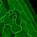
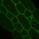
| The cell surface class is characterized by GFP fluorescence that localizes to the extreme periphery of the cell. This pattern is consistent with insertion into or association with the plasma membrane. Plasmolysis tests show that the plasma membrane, not the cell wall, is labeled in these lines. | ||||||||||||||||||||||||||||||||||||||
| Cell surface markers make excellent tools for the visualization of cell shape and patten in two and three dimensions. | ||||||||||||||||||||||||||||||||||||||
 |
||||||||||||||||||||||||||||||||||||||
| Root tip 3D cellular anatomy | ||||||||||||||||||||||||||||||||||||||
 |
||||||||||||||||||||||||||||||||||||||
| Epidermal cell pattern | ||||||||||||||||||||||||||||||||||||||
 |
||||||||||||||||||||||||||||||||||||||
| 3D Guard cell morphology | ||||||||||||||||||||||||||||||||||||||
|
||||||||||||||||||||||||||||||||||||||