| Sub-nuclear localization | |||||||||||||||||||||||||||||||
 |
 |
||||||||||||||||||||||||||||||
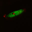
| A single nucleus of a hypocotyl epidermal cell expressing tag 24. This tag displays a fine punctate localization pattern within the nucleus. The red label is plastid autofluorescence. A number of plastids are clustered about the periphery of this nucleus.
A series of flowing dark lines is visible, possibly representing folds and invaginations of the nuclear envelope. One plastid, visible near the center of the nucleus, is associated with this system of dark lines, and appears to be embedded deeply within the nuclear volume. The left set of images are a confocal stack. The right set of images show a 3D brightest point projection of the confocal stack, rotated through 120 degrees. Tag 24 is an out of frame fusion to PIP2A, a plasma membrane water channel. |
|||||||||||||||||||||||||||||||
 |
 |
||||||||||||||||||||||||||||||
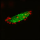
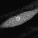
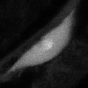
| Tag nucleus (or nuc) accumulates within the nucleolus. Other fusion proteins that loclaize to the nuclear lumen, such as tag 24 above, are excluded from the nucleolus. Outside of the nucleolus, the distribution of tag nuc is relatively uniform.
The images at the left are a confocal stack of a hypocotyl epidermal cell. The second set of images are a 3D brigtest point projection of the confocal stack, rotated through 120 degrees. |
|||||||||||||||||||||||||||||||
|
|||||||||||||||||||||||||||||||