|
|
|
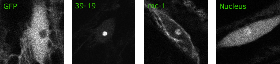
Single confocal sections of hypocotyl cell nuclei showing the diveristy of nucleolar localization patterns. Free GFP is excluded from the nucleolar volume, seen as a dark spherical shadow in the first panel (GFP). We observed three patterns of nucleolar localization: strong nucleolar localization, with little label detected elsewhere in the cell (example: 39-19); localization to both the cytosol and the nucleolus, but significantly reduced elsewhere in the nuclear lumen (example: mc-1), and localization to both the nucleolus and nuclear lumen, but significantly reduced in the cytosol (example: nucleus). |
|