|
|
|
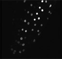 |
3D reconstruction and rotation of a root tip expressing tag M253. Nuclear GFP fluorescence varies greatly among these cells. Part of this variation may be due to the activity of the 35S promotor that is driving expression of this construct, particularly the the relatively greater brightness seen in the outermost layer, the root cap cells, as compared with the second cell layer, the true epidermis. However, the extreme variation seen with cell leyers here is not observed in other nuclear localization lines. There is evidence of a pattern in the variation of cell brightness. "Strings" of cells expressing similar levels of GFP can be seen running along cell files, suggesting the possibility that brightness patterns may be related to clonal cell patterns. |
|