| Root cell mitosis | |||||||||||||||||||||||||||||||
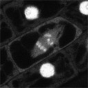 |
Timelapse image series of tag M253 fluorescence in a mitotic root cell. The series begins at prophase, the chromosomes can be seen to line up on a plane at metaphase, and then to separate during anapahse. The number of distinct bodies visualized is consistent with the number of Arabidopsis chromosomes (5). 3-4 of these chomosomes are typically visible on an single confocal plane. Neighboring interphase nuclei are seen as very bright spheres. These nuclei were overexposed slightly to more clearly image the chromosomal fluorescence in the dividing cell.
Note that the cytosolic fluorescence in the dividing cells is relatively higher than that seen in the neighboring cells, suggesting the possibility that some portion of the fusion protein formerly enclosed within the nuclear membrane is now free to diffuse throughout the cytosol. The image series was aquired at a single confcoal plane at 30 second intervals and is played back at 10 frames per second. |
||||||||||||||||||||||||||||||
|
|||||||||||||||||||||||||||||||