|
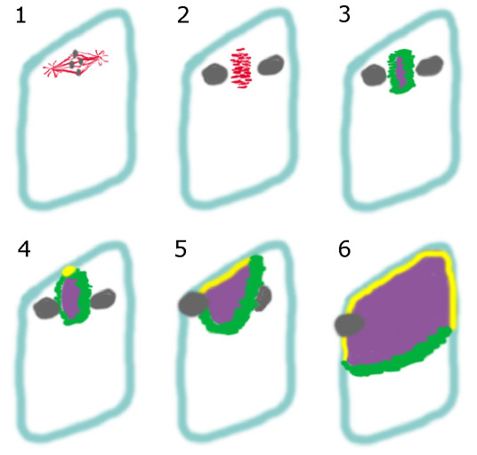
Schematic summary of cytokinetic events observed in vacuolate Arabidopsis cells. The blue line represents the future site of cell plate insertion marked by presumptive cortical cues. 1. The mitotic spindle, shown in red, is adjacent to the cortex of the cell, positioned next to the future site of cell plate insertion. 2. After anaphase, the phragmoplast microtubule raft begins to form midway between the poles of the mitotic spindle. The phragmoplast remains adjacent to the cortex of the cell. 3. Initial elaboration of the phragmoplast (green) and the coalescing cell plate (purple) is symmetrical and grows by radial expansion. 4. Contact of the expanding cell plate occurs at a discreet point along the cortex of the cell, where it contacts with the parental plasma membrane (yellow). 5-6 Two zones of contact between the cell plate and the cortex advance in opposite directions along the marked cortex, building the cell plate in a polarized, directional fashion across the cell body. |
|
|